石墨化碳质物质拉曼光谱温度计原理与应用
Fundamentals and Applications of Raman Spectroscopy of Carbonaceous Material(RSCM)Thermometry
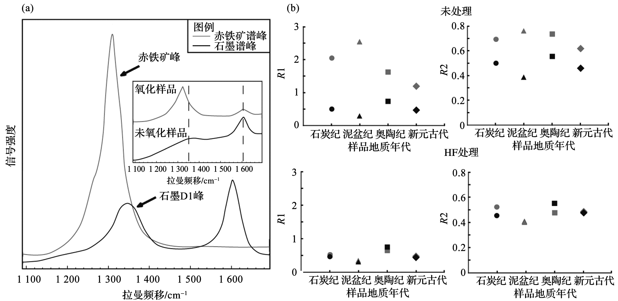
(a)为石墨光谱形态与同频移段内赤铁矿光谱形态,其中赤铁矿谱峰位置为1 320 cm-1,与石墨D1缺陷峰位置(1 350 cm-1)接近,插图为氧化(灰色)与未氧化(黑色)样品的测试谱图,显示氧化样品受赤铁矿谱峰影响而产生虚假的强D1缺陷峰;(b)为是否经HF酸处理的碳质物质R1与R2值的变化:未经过HF处理的氧化样品(灰色)R1与R2值明显大于未氧化样品(黑色);处理后两类样品基本相同
(a) Raman spectra of hematite (grey) and graphite (black). The Raman shift of hematite is located at 1 320 cm-1, close to D1 band (1 350 cm-1) of graphite. The insetted panel shows a comparison of Raman spectra of oxidized (grey) and non-oxidised (black) samples, and the former shows a fake and strong D1 band due to influence of hematite. (b)R1 andR2 plots for untreated and HF-treated samples. Blue points indicate non-oxidised samples. Red points indicate oxidised samples. After treatment, the two types of samples yield similarR1 andR2 values