页岩纳米孔隙研究新进展
张盼盼1,2, 刘小平1,2,*, 王雅杰3, 孙雪娇4
1.中国石油大学(北京)地球科学学院, 北京102249
2.中国石油大学(北京)油气资源与探测国家重点实验室, 北京 102249
3.中石油大港油田分公司勘探开发研究院, 天津 300280
4. 中石油大港油田分公司第五采油厂, 天津 300280
*通讯作者:刘小平(1971-), 男, 安徽无为人, 副教授, 主要从事石油地质教学和研究. E-mail: liuxiaoping@cup.edu.cn

作者简介:张盼盼(1990-), 女, 安徽含山人, 硕士研究生, 主要从事非常规油气地质研究. E-mail: zpp90315@163.com

摘要

随着页岩油气勘探的兴起及近年来北美地区页岩油气开发取得的巨大成功, 含气页岩储层的孔隙研究受到越来越多的重视。页岩储层不同于常规储层, 其以纳米孔隙为主, 无法用常规储层孔隙研究方法进行表征和评价。对目前国内外含气页岩孔隙分类及孔隙表征方法进行了综述, 从定性及定量的角度对表征方法进行归类和总结, 指出了各类方法的优缺点及应用范围。定性表征方法主要是利用聚焦离子束扫描电子显微镜、高分辨率的场发射扫描电子显微镜、透射电子显微镜、宽离子束扫描电子显微镜、原子力显微镜等电子显微成像分析技术及Nano-CT技术等直观描述页岩孔隙的几何形态、连通性和充填情况等;定量表征方法是利用气体吸附法、压汞实验、小角散射及核磁共振等技术定量分析页岩孔径大小及分布、比表面积等。进一步探讨了含气页岩纳米孔隙发育演化的控制因素以及纳米孔隙对页岩气聚集的影响。展望未来, 在页岩纳米孔隙结构表征技术方面, 应不断提高实验精度和效率, 定性与定量表征相结合, 改进三维成像技术;在页岩纳米孔隙储层评价研究方面, 纳米孔隙发育演化的控制因素、纳米孔隙储层的储气及产气能力、陆相非均质页岩纳米孔隙的表征与评价是关注的重点领域。

关键词: 页岩; 孔隙分类; 表征方法; 发育演化; 页岩气聚集
中图分类号:P618.12 文献标志码:A 文章编号:1001-8166(2014)11-1242-08
Research Progress in Shale Nanopores
Zhang Panpan1,2, Liu Xiaoping1,2, Wang Yajie3, Sun Xuejiao4
1. College of Geosciences, China University of Petroleum, Beijing 102249, China
2. State Key Laboratory of etroleum Resources and Prospecting, China University of Petroleum, Beijng 102249, China
3. Research Institute of Petroleum Exploration and Development, Dagang Oilfield Company, CNPC, Tianjin 300280, China
4. The Fifth Oil Production Plant, Dagang Oilfield Company, CNPC, Tianjin 300280, China
Abstract

With the rise of shale oil and gas exploration and great success of North American shale oil and gas development in recent years, the study of gas shale reservoir pores gets more and more attention. The pore diameters of shale reservoirs are mainly in nano-scale which are different from conventional reservoirs, and they can not be evaluated by conventional methods. On the basis of the research on the classification of shale pore types and nanopore characterization techniques, qualitative and quantitative nanopore characterization methods were classified and summarized. In addition, the advantages and disadvantages of various methods as well as application scopes were summarized. Qualitative characterization methods can obtain the morphologic information, connectivity and filling conditions of shale pores directly by focused ion beam milling scanning electron microscopy, field emission scanning electron microscopy, transmission electron microscopy, broad ion beam scanning electron microscope, atomic force microscope and Nano-CT. Quantitative characterization methods can measure pore diameters, pore distribution and specific surface area by nitrogen adsorption experiments, carbon dioxide adsorption experiments, mercury injection experiments, small-angle neutron scattering, and nuclear magnetic resonance. The factors that control nanopore developments and the effects of nanopore on shale gas accumulation were discussed. The nanopore developments are related to total organic carbon content, clay minerals content, carbonate content and thermal maturity. Nanopores have influences on shale gas storage capacity, occurrences of shale gas and seepage mechanisms. Frontier research and development directions were proposed. In the aspect of nanopore characterization techniques of shales, the accuracy and efficiency of the experiments and threedimensional imaging technology should be improved constantly. Qualitative and quantitative characterization techniques should be combined. As to the evaluation of shale nanopore reservoirs, the factors that control nanopore developments, shale gas storing and producing and nanopores characterization and evaluation of continental heterogeneous shale are the key issues for further research.

Keyword: Shale; Pore classification; Characterization methods; Developmental evolution; Shale gas gathering.
1 引言

近年来, 随着北美地区页岩气成功勘探开发, 页岩气已成为世界非常规资源勘探开发的热点。页岩气主要以吸附和游离状态赋存于页岩中, 吸附态页岩气存在于有机质和黏土矿物表面, 游离态页岩气存在于孔隙和裂隙中, 还有少量溶解于液态烃和水中的溶解态页岩气。因此, 页岩不仅能作为烃源岩和盖层, 还能成为储层。页岩作为一种超致密油气储层, 其孔隙远远小于常规的砂岩储层和碳酸盐储层, 孔径大小达到纳米量级[1]。储层岩石的孔隙结构是影响天然气储集能力和页岩气开采的主要因素[2]。Chalmers等[3]研究发现Haynesvil页岩、Woodford页岩、Marcellus页岩和Barnett页岩的平均孔隙直径分别为4.9, 5.5, 3.9和4.0 nm, 指出页岩纳米孔隙结构控制页岩油气储集能力及裂缝网络系统的流体运移能力。中国四川盆地南部寒武系、志留系等高成熟页岩孔隙直径以150 nm为主[4], 在马朗凹陷芦草沟组页岩中也发现了丰富的纳米级有机质孔[5]

页岩储层中纳米孔隙的发现, 标志着储层孔隙结构研究重点已从常规储层的毫米至微米级孔隙转向非常规储层的纳米级孔隙。纳米孔隙结构的表征、纳米孔隙发育规律及纳米孔隙对页岩油气聚集的影响等是研究的重点和难点, 其对页岩油气资源评价以及勘探开发都具有重要意义。

2 页岩孔隙分类

国外学者主要从孔径的大小、孔隙赋存状态及孔隙的连通性对页岩孔隙进行划分。目前提出的页岩储层孔隙大小的分类方案中[6, 7, 8, 9, 10, 11, 12, 13, 14, 15], 应用最广的是国际理论和应用化学学会(International Union of Pure and Applied Chemistry, 简称IUPAC)的分类方案。根据IUPAC的分类[9, 10], 孔径小于2 nm的孔隙为微孔(Micropore), 孔径在2~50 nm的孔隙为介孔(Mesopore), 大于50 nm的孔隙为大孔(Macropore)。根据孔隙的赋存状态可以将纳米孔隙分为粒内孔、粒间孔和有机质孔[11]。根据孔隙的连通性将孔隙分为开孔和闭孔, 开孔进一步可分为盲孔和通孔[1]

以上分类方案中缺少对孔隙有效性的考虑, 孔隙有效性主要是含油气能力大小及对渗流的影响等。由于页岩独特的纳米孔隙结构及复杂的油气赋存状态, 导致油气在孔隙中渗流机理更加复杂, 因此纳米孔隙含油气性仍是研究中的难题。加强纳米孔隙内流体渗流特征模拟与研究, 对分析纳米孔隙有效性具有重要意义。

3 页岩纳米孔隙研究方法与表征

页岩储层不同于常规储层, 具有孔径小、孔隙度渗透率低等特点, 无法用常规的储层孔隙评价方法进行表征。近年来, 石油工业采用聚焦离子束扫描电子显微镜(Focused Ion Beam Scanning Electron Microscopy, 简称FIB-SEM)、高分辨率的场发射扫描电子显微镜(Field Emission Scanning Electron Microscopy, 简称FE-SEM)、透射电子显微镜(Transmission Electron Microscopy, 简称TEM)、宽离子束扫描电子显微镜(Broad Ion Beam Scanning Electron Microscopy, 简称BIB-SEM)、原子力显微镜(Atomic Force Microscopy, 简称AFM)等电子显微成像技术以及Nano-CT、能谱仪(Energy Dispersive Spectrometer, 简称EDS)、高压压汞(Mercury Injection Capillary Pressure, 简称MICP)、低压N2和CO2吸附实验、核磁共振光谱(Nuclear Magnetic Resonance, 简称NMR)、小角散射(Small-Angle Scattering, 简称SAS)等一系列先进技术来研究页岩纳米孔隙结构[3, 15, 16, 17, 18, 19, 20, 21, 22, 23, 24, 25, 26, 27, 28, 29, 30, 31, 32](图1)。对页岩纳米孔隙几何形态、连通性、充填情况及大小的直观观测, 主要是利用电子显微镜成像分析、Nano-CT等技术进行定性分析; 对纳米孔径大小分布、比表面积的定量分析主要借助于MICP、气体吸附、NMR、SAS等技术。

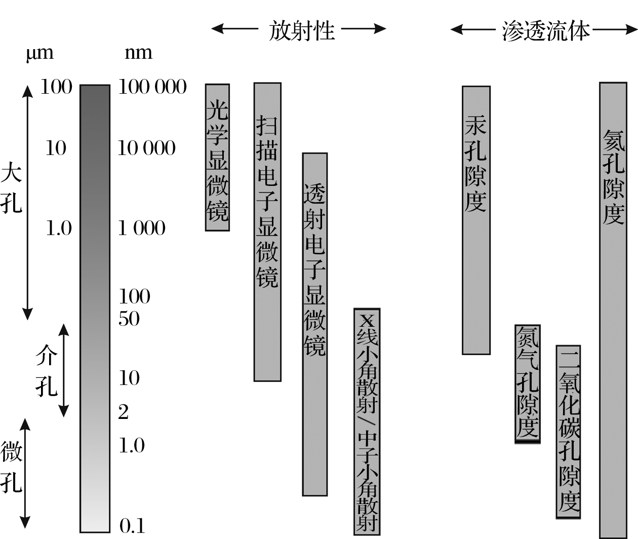
图1 表征纳米孔隙研究方法的应用范围[32]Fig.2 Utility of various methodologies in common use for investing porous materials[32]

3.1 定性表征方法

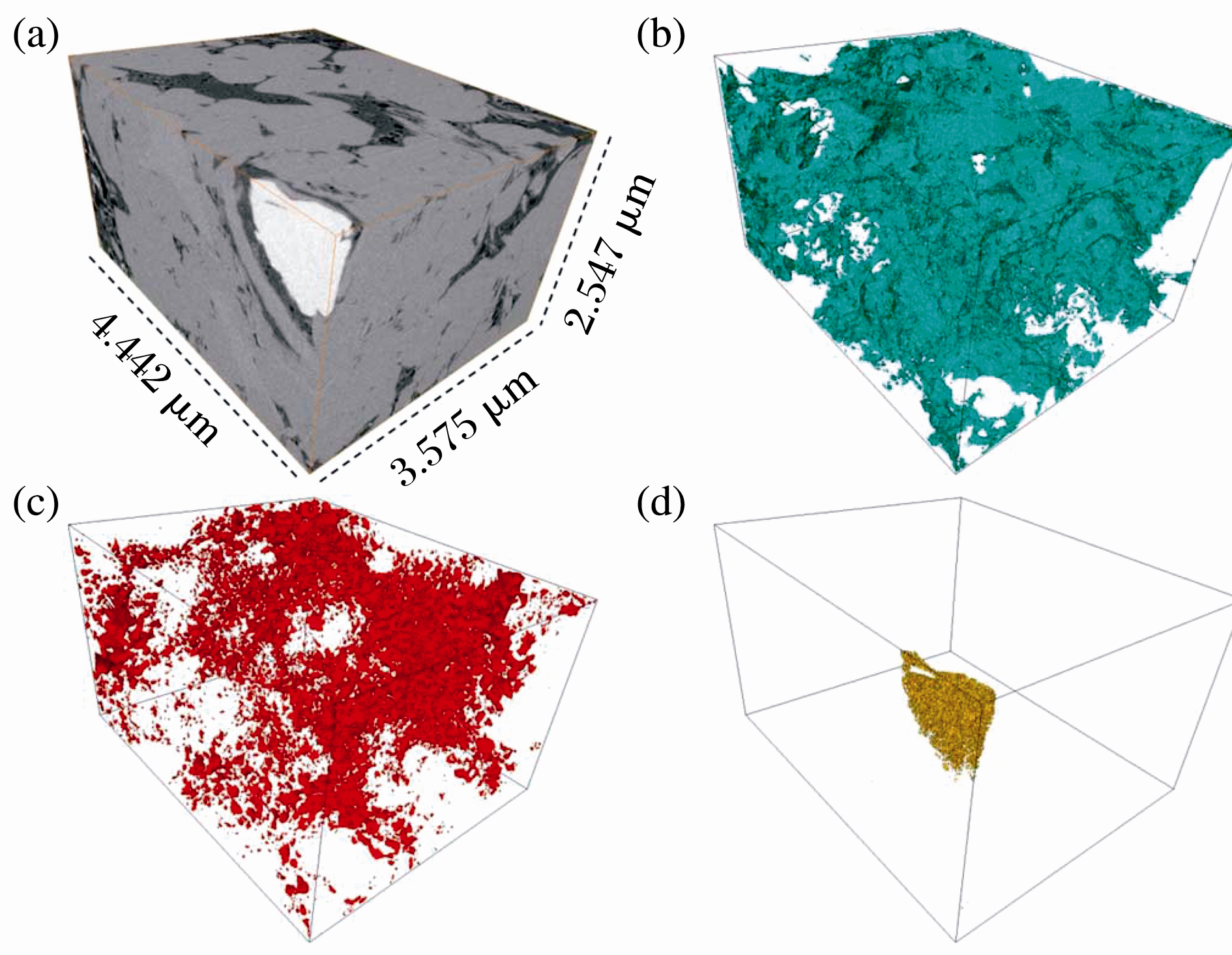
对页岩纳米孔隙几何形态、大小及连通性的定性观察主要是利用SEM、AFM、TEM、FE-SEM、FIB-SEM等微区观察技术, 精确度受样品本身、样品处理方式及仪器的分辨率控制。在对样品进行预处理时, 需要利用氩离子抛光技术替代机械抛光, 利于显微镜下的观察。SEM是一种利用电子束扫描样品表面从而获得样品信息的电子显微镜, 由于页岩薄片不规则的表面形态, 不能用来观察介孔和微孔[2, 3, 15, 16]。AFM通过检测样品表面和一个微型力敏感元件之间的极微弱的原子间相互作用力来研究样品的表面结构及性质, 主要用来观察页岩纳米孔隙的介孔[17, 18]。AFM可在常压环境下工作, 不需要对样品进行特殊处理, 并可提供孔隙的三维图像, 缺点在于成像范围小, 速度慢, 受探头的影响大[17, 18]。TEM是一种根据电子穿过介质作用的成像技术, 可提供介孔和大孔的三维孔隙图, 分辨率下限可达0.2 nm[3, 19]。FE-SEM利用反映表面形貌结构的二次电子信号与反射成分信息的背散射电子信号, 结合X射线能谱仪测量孔隙半径、喉道宽度等, 观测精度可达0.1 nm [3, 33](图2)。FIB-SEM 是将聚焦离子束光刻技术(FIB milling)与扫描电子显微镜成像相结合, 实现切割与扫描同时进行, 提供的三维图像可更清楚地研究纳米孔隙的分布及连通性(图3)[25]。FIB-SEM的缺点在于制样时损坏了样品表面, 成像范围太小。国外学者[26]提出宽离子束光刻和扫描电子显微镜相结合的方法(BIB-SEM), 弥补FIB-SEM的不足。Curtis等[25]利用FIB-SEM, 并结合背散射电子图像(BSE)及能量色散谱观察比较Barnett, Eagle Ford, Fayetteville, Floyd等9个地层页岩样品的微观结构, 在干酪根和无机质中都发现了纳米孔隙。Nano-CT技术可建立不同尺度孔喉三维空间模型, 实现孔喉连通性评价研究。其分辨率可达到50~70 nm, 具有不损坏样品的独到优势, 但在实验过程中受到测试环境及页岩非均质性等方面的影响[1]。邹才能等[4]利用场发射扫描电子显微镜与Nano-CT重构技术, 在四川盆地南部古生界高成熟页岩中发现了丰富的纳米级孔隙。

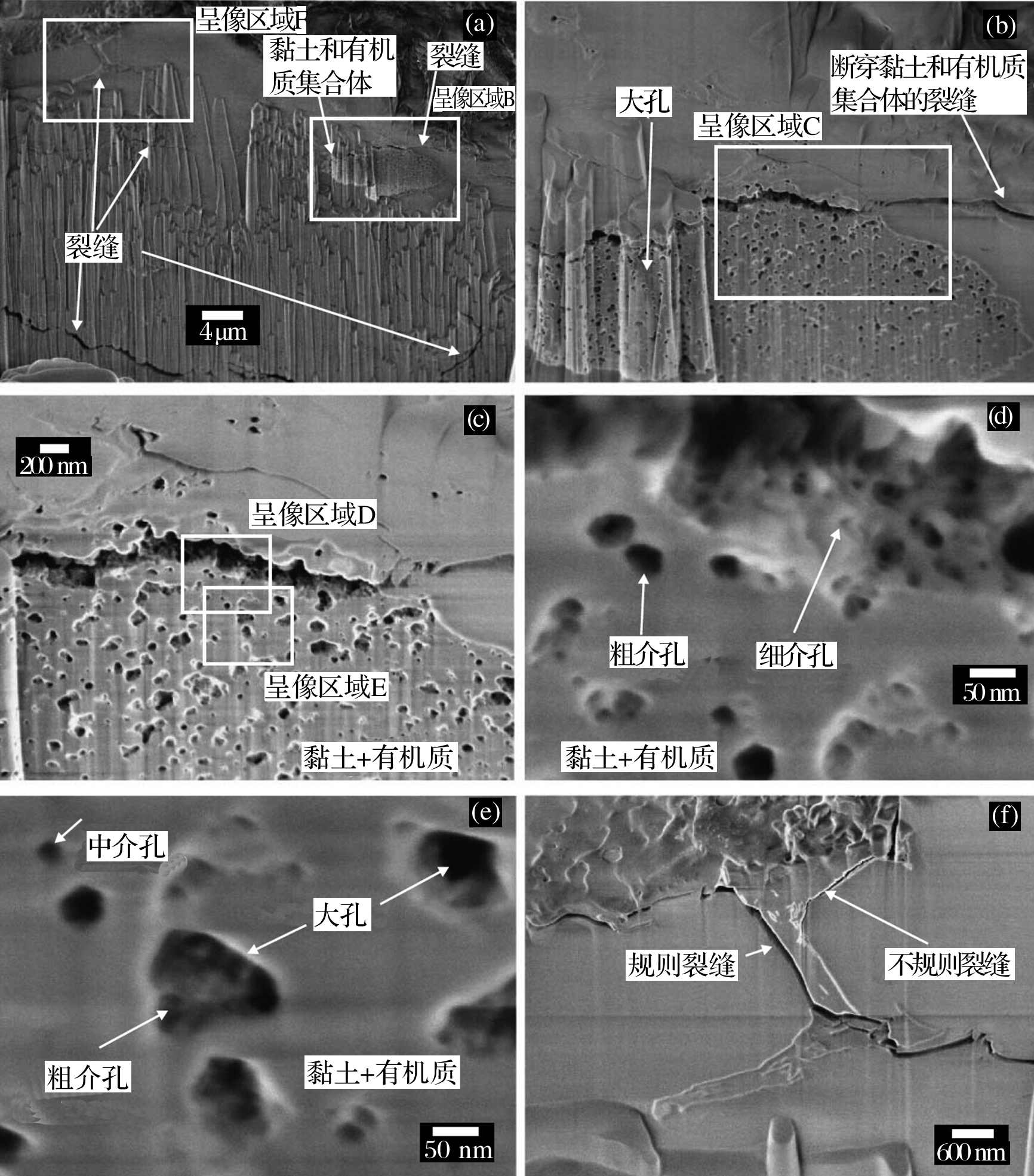
图2 利用FE-SEM观察Woodford页岩样品[3]Fig.2 Field emission scanning electron microscope(FE-SEM) images of the Woodford sample[3]

图3 利用FIB-SEM背散射电子图像重构Horn River页岩样品三维图[25](a)三维立体图; (b)干酪根; (c)孔隙; (d)黄铁矿Fig.3 Reconstruction of the Horn River sample from 500 sequential backscattered electron images showing[25](a)A three-dimensional solid; (b) Kerogen; (c)Pores; (d) Pyrite

3.2 定量表征方法

MICP、低压N2吸附、低压CO2吸附、He孔隙率测定、NMR、SAS等技术可定量表征页岩纳米孔隙孔径大小分布及比表面积。MICP的测试下限较大, 主要用于测定大孔(> 50 nm)分布特征及孔隙度, 利用BET方程和BJH方程处理N2吸附数据可得到比表面积和介孔(2~50 nm)孔径分布特征, 通过Dubinin-Radushkevich(D-R)理论模型处理CO2吸附数据可得到微孔(< 2 nm)的分布特征[1, 3]。为了更好的描述孔径分布, 可以将压汞过程中对渗透率起明显贡献作用的最小孔径作为压汞测试法描述的孔径下限, 而低于此下限的孔隙再利用N2吸附和CO2吸附分别测定。目前这些方法是研究页岩孔隙度、孔径分布、比表面积等特征的重要工具, 但也存在着一定局限性。首先, 这些方法仅限于研究开孔, 不能表征闭孔, 实验结果会受样品粒度影响, 样品粒度越小, 闭孔就有可能转变为开孔, 导致开孔数目的增加, 使所测的比表面积及孔隙度变大; 在页岩样品预热过程中, 水分的散失会产生裂隙, 要充分考虑样品粒度及含水性的影响。在进行压汞法测试时, 高压会导致页岩结构的破坏, 影响孔隙测量结果; 此外, 孔径分布的计算式基于Washburn方程, 而Washburn方程是假设样品孔隙为光滑的圆柱状孔隙, 页岩纳米孔隙的复杂性会对结果造成误差; 压汞存在的瓶颈效应及边界效应可能会对孔径分布结果造成影响[1]

NMR研究孔隙主要基于弛豫, 尤其是表面弛豫对孔隙结构的灵敏反应, 其能在基本不受岩石骨架成分影响下获得岩层孔隙度及孔隙流体等信息[19, 25, 34]。但核磁共振光谱学法孔隙度受测试环境、仪器参数、样品的微孔隙、顺磁性物质及流体类型等多种因素影响[23]。小角散射技术(SAS)主要包括中子小角散射(Small-Angle Neutron Scattering, SANS)和X线小角散射(Small-Angle X-Ray Scattering, SANS SAXS), 其与超小角散射(Ultra-Small-Angle Scattering, USAS)的工作原理一致, 利用中子射线或者X射线照射样品, 通过散射辐射的强度与散射角之间的关系反映样品微结构信息[27, 28, 30]。中子超小角散射(Ultra-Small-Angle Neutron Scattering, USANS)的测试范围在100 nm~10 μ m之间, 而SANS的测试孔径< 250 nm, X线小角散射观察孔径下限为0.2 nm[27]。SAS及USAS的优势在于处理样品时对样品无损坏, 并可在不同温压条件下研究样品; 它们还可以表征页岩储层的闭孔及判断孔隙的含气性[28]。但是, 由于页岩非均质性强的特点, 会导致核散射界面、电子密度差异使得实验结果出现较大误差, 此外研究所需要的中子源数量稀少, 研究费用昂贵。

4 影响页岩纳米孔隙发育的因素

页岩储层纳米孔隙的发育受多种因素共同控制。国内外学者通过研究认为其与页岩总有机碳含量(TOC)、黏土矿物含量、碳酸盐含量及有机质热演化存在对应关系 [35, 36, 37, 38, 39, 40, 41]。有机碳含量与比表面积总孔体积成正相关关系, 且是控制纳米级孔隙体积及其比表面积的主要内在因素[3, 41]。有机质颗粒内纳米孔隙主要是由于生成液体或气体聚积生成气泡而成, 其富集和形成与有机质的成熟生烃有关, 并形成有效的孔隙网络, 主导油气渗流通道[2, 13, 15, 42]。Jarvie等[43]指出页岩中孔隙以有机质纳米孔隙为主, 随着成熟度的增大而增加, 其形成和演化可分成3个阶段:形成期(0.6%< Ro< 2.0%)、发展期(2.0%< Ro< 3.5%)和转换破坏期(> 3.5%)。Milliken等[44]利用FE-SEM对宾夕法尼亚(Pennsylvania)的Marcellus页岩储层进行研究, 认为TOC对有机质纳米孔的影响超过与成熟度对其的影响, 当TOC< 5.5%时, TOC与有机质孔具有正相关关系, 当TOC超过5.5%时, 其与有机质孔的相关性不明显。除了页岩的内在因素外, 超压以及有机酸引起的溶蚀作用等也是影响纳米孔隙的发育的重要因素。从沉积开始到压实、成岩、生烃与排烃等过程中, 页岩孔隙主体演化趋势是由大变小, 最终保持稳定 [45]。而超压的形成在一定程度上减缓了孔隙度变小的速率, 有利于纳米孔隙的保存。超压的成因很多, 生烃作用和差异压实作用是最主要的两种成因[46, 47, 48]。此外, 有研究表明温度和压力在一定程度上控制低成熟页岩孔隙发育[49]

5 纳米孔隙对页岩气聚集的影响

页岩储层特殊的纳米孔隙结构影响着页岩气的聚集, 主要体现在页岩储气能力、页岩气赋存形式及渗流机理3个方面[50]。页岩气的赋存形式具有多样性, 包括游离态、吸附态及溶解态, 但以游离态和吸附态为主[42, 51, 52]。页岩纳米孔隙结构的大孔和介孔有利于游离态页岩气的储存, 微孔和部分介孔则有利于吸附气的储存[53]。因为相对于大孔和介孔而言, 微孔的总体积越大, 其比表面积越大, 对气体分子的吸附能力也就越强[51]。孔隙度是决定页岩的气体总量的因素之一, 与页岩气体总量呈正相关关系, 即页岩的气体总含量随着页岩孔隙度的增大而增大[54]。页岩的吸附含气量受多重因素影响, 纳米孔隙体积、比表面积、有机碳含量与吸附气含量呈正相关关系[41, 55]。较高的微孔虽能保证页岩储层具有很高的吸附聚气能力, 但若介孔和大孔发育较差, 则不利于气体渗流和页岩气的开发[56]。此外, 由于纳米孔隙的大量存在, 且与微米级孔隙、微裂缝相连接的孔隙网络及吸附气大量存在, 导致气藏同时存在渗流和扩散, 使流动机理更加复杂。气体的传质方式也受孔隙尺度的影响, 在气藏开采初期和中期, 直径> 10 nm的孔隙是主要传质通道[57]。纳米孔隙特征还影响着页岩基质的渗透率[3]。纳米孔隙的大量存在使得页岩储层在早期认识上属于不渗或超低渗的范畴, 但生产数据表明页岩气的产气能力并不低, 且页岩生产中的表观渗透率与达西渗透率的比值随着孔径减小而增大, 说明纳米孔隙内气体分子扩散及滑脱作用对气体渗流影响很大[58]。此外对其深入研究, 有利于对储层后期改造研究[59]

6 展望

页岩储层独特的纳米孔隙结构, 增加了页岩储层的评价难度。尽管国内外学者针对页岩纳米孔隙结构提出了多种研究方法, 但是由于仪器本身分辨率的限制及研究方法的局限性, 目前, 还没有一套精准的用来综合评价纳米孔隙的方法。在页岩储层纳米孔隙结构的表征技术方面, 未来研究的发展趋势是:①提高实验精度和效率。进一步研发新方法和新仪器, 使得纳米孔隙实验研究结果精度更高, 同时也更加便捷、价格低廉及样品低损耗。②定性与定量表征相结合。任何一种实验技术和方法都存在一定的局限性, 应将定性与定量方法相结合, 从而建立有效的表征方法组合。③改进纳米孔隙的三维成像技术, 扩大成像范围。三维成像技术的不断改进可以更加有效地研究不同尺度、不同类型纳米孔隙结构的连通性及含油气性, 提高对非均质性页岩储层表征的系统性和代表性。

在页岩纳米孔隙储层评价研究方面, 未来重点关注的领域主要为:①纳米孔隙发育演化的控制因素研究。影响纳米孔隙发育演化的内在和外在因素多, 目前的研究主要集中于单因素分析, 尚缺乏系统性和多因素综合分析。②页岩纳米孔隙储层的储气及产气能力研究。页岩纳米孔隙结构及其发育的非均质性对页岩储气能力、页岩气赋存形式及渗流机理的影响是今后研究的重点。③陆相非均质页岩纳米孔隙的表征与评价。我国广泛发育湖相页岩, 由于海相与陆相页岩储层的差异性, 在借鉴北美海相含气页岩储层纳米孔隙结构评价方法和标准的同时, 要加强研究陆相页岩的非均质性及其纳米孔隙结构特点, 建立符合陆相页岩储层特点的纳米孔隙研究方法体系, 为陆相页岩油气资源评价及勘探开发提供理论基础。

The authors have declared that no competing interests exist.

参考文献
[1] Cui Jingwei, Zou Caineng, Zhu Rukai, et al. New advances in shale porosity research[J]. Advances in Earth Science, 2012, 27(12): 1319-1325.
[崔景伟, 邹才能, 朱如凯, . 页岩孔隙研究新进展[J]. 地球科学进展, 2012, 27(12): 1319-1325. ] [本文引用:4] [CJCR: 1.388]
[2] Ambrose R J, Hartman R C, Diaz-Campos M, et al. New pore-scale considerations for shale gas in place calculations[C]\\ SPE Unconventional Gas Conference. Pittsburgh, Pennsylvania, USA, 2010. [本文引用:3]
[3] Chalmers G R, Bustin R M, Power I M. Characterization of gas shale pore systems by porosimetry, pycnometry, surface area, and field emission scanning electron microscopy/transmission electron microscopy image analyses: Examples from the Barnett, Woodford, Haynesville, Marcellus, and Doig units[J]. AAPG Bulletin, 2012, 96(6): 1099-1119. [本文引用:8] [JCR: 1.768]
[4] Zou Caineng, Zhu Rukai, Bai Bin, et al. First discovery of nano-pore throat in oil and gas reservoir in China and its scientific value[J]. Acta Petrologica Sinica, 2011, 27(6): 1857-1864.
[邹才能, 朱如凯, 白斌, . 中国油气储层中纳米孔首次发现机及其科学价值[J]. 岩石学报, 2011, 27(6): 1857-1864. ] [本文引用:2] [JCR: 1.117] [CJCR: 2.65]
[5] Huang Zhilong, Guo Xiaobo, Liu Bo, et al. The reservoir space characteristics and origins of Lucaogou Formation source rock oil in the Malang Sag[J]. Acta Sedimentologica Sinica, 2012, 30(6): 1115-1122.
[黄志龙, 郭小波, 柳波, . 马朗凹陷芦草沟组源岩油储集空间特征及其成因[J]. 沉积学报, 2012, 30(6): 1115-1122. ] [本文引用:1] [CJCR: 1.227]
[6] Zou Caineng, Tao Shizhen, Hou Lianhua, et al. Unconventional Petroleum Geology[M]. Beijing: Geology Publishing House, 2011.
[邹才能, 陶士振, 侯连华, . 非常规油气地质[M]. 北京: 地质出版社, 2011. ] [本文引用:1]
[7] American Society for TestingMaterials. ASTM D1412-07 Stand ard Test Method for Equilibrium Moisture of Coal at 96 to 97 Percent Relative Humidity and 30 ℃[S]. West Conshohocken: ASTM International, 2004: 261-263. [本文引用:1]
[8] Ambrose R J, Hartman R C, Diaz-Campos M. et al. Shale gas-in-place calculations PartⅠ: New pore scale consideration[J]. SPE Journal, 2012, 17(1): 219-229. [本文引用:1] [JCR: 1.011]
[9] Gregg S J, Sing K S W. Adsorption, Surface Area and Porosity[M]. London: Academic Press, 1982. [本文引用:2]
[10] Sing K S W, Everett D H, Haul R A W, et al. Reporting physisorption data for gas/solid systems with special reference to the determination of surface area and porosity[J]. Pure and Applied Chemistry, 1985, 57(4): 603-619. [本文引用:2] [JCR: 3.386]
[11] Loucks R G, Reed R M, Ruppel S C. Spectrum of pore types and networks in mudrocks and a descriptive classification for matrix-related mudrock pores[J]. AAPG Bulletin, 2012, 96(6): 1071-1098. [本文引用:2] [JCR: 1.768]
[12] Desbois G, Urai J L, Kukla P A. Morphology of the pore space in claystones—Evidence from BIB/FIB ion beam sectioning and cryo-SEM observations[J]. Earth, 2009, 4: 15-22. [本文引用:1]
[13] Loucks R G, Reed R M, Ruppel S C, et al. Morphology, genesis, and distribution of nanometer-scale pores in siliceous mudstones of the Mississippian Barnett Shale[J]. Journal of Sedimentary Research, 2009, 79(11/12): 848-861. [本文引用:2] [JCR: 1.742]
[14] Slatt E M, O’Neal N R. Pore types in the Barnett and Woodford gas shales: Contribution to understand ing gas storage and migration pathways in fine-grained rocks[J]. AAPG Bulletin, 2011, 95(12): 2017-2030. [本文引用:1] [JCR: 1.768]
[15] Curtis M E, Ambrose R J, Sondergeld C H, et al. Structural characterization of gas shales on the micro- and nano-scales[C]\\ SPE Canadian Unconventional Resources and International Petroleum Conference. Calgary, Alberta, Canada, 2010. [本文引用:4]
[16] Sondergeld C H, Ambrose R J, Rai C S, et al. Microstructural studies of gas shales[C]\\ SPE Unconventional Gas Conference. Pittsburgh, Pennsylvania, USA, 2010. [本文引用:2]
[17] Javadpour F. Nanopores and apparent permeability of gas flow in mudrocks(shales and siltstone)[J]. Journal of Canadian Petroleum Technology, 2009, 48(8): 16-21. [本文引用:3] [JCR: 0.451]
[18] Javadpour F, Moravvej Farshi M, Amrein M. Atomic-force microscopy: A new tool for gas-shale characterization[J]. Journal of Canadian Petroleum Technology, 2012, 51(4): 236-243. [本文引用:3] [JCR: 0.451]
[19] Curtis M E, Ambrose R J, Sondergeld C H, et al. Transmission and scanning electron microscopy investigation of pore connectivity of gas shales on the nanoscale[C]\\ SPE Unconventional Gas Conference. Woodland s, Texas, USA, 2011. [本文引用:3]
[20] Milner M, Mclin R, Petrirllo J, et al. Imaging texture and porosity in mudstones and shales: Comparison of secondary and ion-milled backscatter SEM methods[C]\\ SPE Unconventional Resources & International Petroleum Conference. Calgary, Alberta, Canada, 2010. [本文引用:1]
[21] Sondergeld C H, Ambrose R J, Rai C S, et al. Micro-structural studies of gas shales[C]\\ SPE Unconventional Gas Conference. Pittsburgh, Pennsyvania, USA, 2010. [本文引用:1]
[22] Farshi M M, Javadpour F. Uncovering nanoscale issues in shale gas systems[J]. Geological Society of America, 2011, 43(3): 48. [本文引用:1] [JCR: 4.286]
[23] Bustin A M M, Bustin R M, Cui X. Importance of fabric on the production of gas shales[C]\\ SPE Unconventional Reservoirs Conference. Keystone, Colorado, USA, 2008. [本文引用:2]
[24] Golab A N, Knackstedt M A, Averdunk H, et al. 3D porosity and mineralogy characterization in tight gas sand stones[J]. Special Section: Tight Gas Sand s, 2010, 29(12): 1476-1483. [本文引用:1]
[25] Curtis M E, Sondergeld C H, Ambrose R J, et al. Microstructural investigation of gas shales in two and three dimensions using nanometer-scale resolution imaging[J]. AAPG Bulletin, 2012, 96(4): 665-677. [本文引用:4] [JCR: 1.768]
[26] Klaver J, Desbois G, Urai J L, et al. BIB-SEM study of the pore space morphology in early mature Posidonia Shale from the Hils area, Germany[J]. International Journal of Coal Geology, 2012, 103: 12-25. [本文引用:2] [JCR: 2.976]
[27] Ruppert L F, Sakurovs R, Blach T P, et al. A USANS/SANS study of the accessibility of pores in the Barnett Shale to methane and water[J]. Energy Fuels, 2013, 27: 772-779. [本文引用:3] [JCR: 2.853]
[28] Clarkson C R, Freeman M, He L, et al. Characterization of tight gas reservoir pore structure using USANS/SANS and gas adsorption analysis[J]. Fuel, 2012, 95: 371-385. [本文引用:3] [JCR: 3.357]
[29] Clarkson C R, Solano N, Bustin R M, et al. Pore structure characterization of North American shale gas reservoirs using USANS/SANS, gas adsorption, and mercury intrusion[J]. Fuel, 2013, 103: 606-616. [本文引用:1] [JCR: 3.357]
[30] Melnichenko Y B, He L, Sakurovs R, et al. Accessibility of pores in coal to methane and carbon dioxide[J]. Fuel, 2012, 91: 200-208. [本文引用:2] [JCR: 3.357]
[31] Sigal R F, Odusina E. Laboratory NMR measurements on methane saturated Barnett Shale samples[J]. Petrophysics, 2011, 52: 32-49. [本文引用:1] [JCR: 0.4]
[32] Bustin R M, Bustin A M M, Cui X, et al. Impact of shale properties on pore structure and storage characterisitics[C]\\ SPE Society of Petroleum Engineers. Fort Worth, Texas, USA, 2008. [本文引用:1]
[33] Bai Bin, Zhu Rukai, Wu Songtao, et al. New micro-throat structural characterization techniques for unconventional tight hydrocarbon reservoir[J]. China Petroleum Exploration, 2014, 19(3): 78-86.
[白斌, 朱如凯, 吴松涛, . 非常规油气致密储层微观孔喉结构表征新技术及意义[J]. 中国石油勘探, 2014, 19(3): 78-86. ] [本文引用:1] [CJCR: 0.8848]
[34] Kleiberg R L, Kenyon W E, Mitra P P. Mechanism of NMR relaxation of fluids in rock[J]. Journal of Magnetic Resonance(Series A), 1994, 108(2): 206-214. [本文引用:1]
[35] Kuila U, Prasad M. Surface area and pore-size distribution in clays and shales[J]. Geophysical Prospecting, 2013, 61(2): 341-362. [本文引用:1] [JCR: 1.36]
[36] Modica C J, Lapierre S G. Estimation of kerogen porosity in source rocks as a function of thermal transformation: Example from the Mowry Shale in the Powder River Basin of Wyoming[J]. AAPG Bulletin, 2012, 96(1): 87-108. [本文引用:1] [JCR: 1.768]
[37] Chalmers G R, Bustin R M. Light volatile liquid and gas shale reservoir potential of the Cretaceous Shaftesbury Formation in northeastern British Columbia, Canada[J]. AAPG Bulletin, 2012, 96(7): 1333-1367. [本文引用:1] [JCR: 1.768]
[38] Curtis M E, Cardott B J, Sondergeld C H, et al. Development of organic porosity in the Woodford Shale with increasing thermal maturity[J]. International Journal of Coal Geology, 2012, 103: 26-31. [本文引用:1] [JCR: 2.976]
[39] Romero-Sarmiento M F, Rouzaud J N, Bernard S, et al. Evolution of Barnett Shale organic carbon structure and nanostructure with increasing maturation[J]. Organic Geochemistry, 2014, 71: 7-16. [本文引用:1] [JCR: 2.518]
[40] Rose D J K, Bustin R M. The importance of shale composition and pore structure upon gas storage potential of shale gas reservoirs[J]. Marine and Petroleum Geology, 2009, 26(6): 916-927. [本文引用:1] [JCR: 2.111]
[41] Han Shuangbiao, Zhang Jinchuan, Yang Chao, et al. The characteristics of nanoscale pore and its gas storage capability in the Lower Cambrian shale of southeast Chongqing[J]. Journal of China Coal Society, 2013, 38(6): 1038-1043.
[韩双彪, 张金川, 杨超, . 渝东南下寒武页岩纳米孔隙特征及其储气性能[J]. 煤炭学报, 2013, 38(6): 1038-1043. ] [本文引用:3] [CJCR: 0.787]
[42] Javadpour F, Fisher D, Unsworth M. Nanoscale gas flow in shale gas sediments[J]. Journal of Canadian Petroleum Technology, 2007, 46(10): 55-61. [本文引用:2] [JCR: 0.451]
[43] Jarvie D M, Hill R J, Ruble T E, et al. Unconventional shale-gas systems: The Mississippian Barnett Shale of north-central Texas as one model for thermogenic shale-gas assessment[J]. AAPG Bulletin, 2007, 91(4): 475-799. [本文引用:1] [JCR: 1.768]
[44] Milliken K L, Rudnicki M, Awwiller D N, et al. Organic matter-hosted pore system, Marcellus Formation (Devonian), Pennsylvania[J]. AAPG Bulletin, 2013, 97(2): 177-200. [本文引用:1] [JCR: 1.768]
[45] Guo Qiulin, Chen Xiaoming, Song Huanqi, et al. Evolution and models of shale porosity during burial process[J]. Natural Gas Geoscience, 2013, 24(3): 439-449.
[郭秋麟, 陈晓明, 宋焕琪, . 泥页岩埋藏过程孔隙度演化与预测模型探讨[J]. 天然气地球科学, 2013, 24(3): 439-449. ] [本文引用:1] [CJCR: 1.055]
[46] Li Mingcheng. Oil and Gas Migration[M]. Beijing: Petroleum Industry Press, 2004: 50-57.
[李明诚. 石油与天然气运移[M]. 北京: 石油工业出版社, 2004: 50-57. ] [本文引用:1]
[47] Guo Qiulin, Li Jianzhong, Chen Ningsheng, et al. Modeling of the tight sand stone gas accumulation for the Xujiahe Formation, Hechuan-Tongnan area, Sichuan Basin[J]. Petroleum Exploration and Development, 2011, 38(4): 409-417.
[郭秋麟, 李建忠, 陈宁生, . 四川合川—潼南地区须家河组致密砂岩气成藏模拟[J]. 石油勘探开发, 2011, 38(4): 409-417. ] [本文引用:1] [CJCR: 2.573]
[48] Guo Qiulin, Chen Ningsheng, Hu Junwen, et al. Geo-model of tight sand stone gas accumulation and quantitative simulation[J]. Natural Gas Geoscience, 2012, 23(2): 199-207.
[郭秋麟, 陈宁生, 胡俊文, . 致密砂岩气聚集模型与定量模拟探讨[J]. 天然气地球科学, 2012, 23(2): 199-207. ] [本文引用:1] [CJCR: 1.055]
[49] Zhu Rukai, Bai Bin, Cui Jingwei, et al. Research advances of microstructure in unconventional tight oil and gas reservoitrs[J]. Journal of Palaeogeography, 2013, 15(5): 615-623.
[朱如凯, 白斌, 崔景伟, . 非常规油气致密储集层微观结构研究进展[J]. 古地理学报, 2013, 15(5): 615-623. ] [本文引用:1]
[50] Zeng Jianhui, Yang Zhifeng, Feng Xiao, et al. Study status and key scientific issue of tight reservoir oil and gas accumulation mechanism[J]. Advances in Earth Science, 2014, 29(6): 651-661.
[曾溅辉, 杨智峰, 冯枭, . 致密储层油气成藏机理研究现状及其关键科学问题[J]. 地球科学进展, 2014, 29(6): 651-661. ] [本文引用:1] [CJCR: 1.388]
[51] Zhang Xuefen, Lu Xiancai, Zhang Linye, et al. Occurrences of shale gas and their petroleum geological significance[J]. Advances in Earth Science, 2010, 25(6) : 597-604.
[张雪芬, 陆现彩, 张林晔, . 页岩气的赋存形式研究及其石油地质意义[J]. 地球科学进展, 2010, 25(6): 597-604. ] [本文引用:2] [CJCR: 1.388]
[52] Etminan S R, Javadpour F, Maini B B, et al. Measurement of gas storage processes in shale and of the molecular diffusion coefficient in kerogen[J]. International Journal of Coal Geology, 2014, 123: 10-19. [本文引用:1] [JCR: 2.976]
[53] Ross D J K, Bustin R M. Impact of mass balance calculations on adsorption capacities in microporous shale gas reservoirs[J]. Original Research Article Fuel, 2007, 86(17/18): 2696-2706. [本文引用:1]
[54] Chalmers G R L, Bustin R M. Lower Cretaceous gas shales in northeastern British Columbia, evaluation of regional potential gas resource[J]. Bulletin of Canadian Petroleum Geology, 2008, 56(1): 22-61. [本文引用:1] [JCR: 0.806]
[55] Chalmers G R L, Bustin R M. The organic matter distribution and methane capacity of the Lower Cretaceous strata of Northeastern British Columbia, Canada[J]. International Journal of Coal Geology, 2007, 70: 223-239. [本文引用:1] [JCR: 2.976]
[56] Yang Feng, Ning Zhengfu, Kong Detao, et al. Pore structures of shales from high pressure mercury injection and nitrogen adsorption methond[J]. Natural Gas Geoscience, 2013, 24(3): 450-455.
[杨峰, 宁正福, 孔德涛, . 高压压汞法和氮气吸附法分析页岩孔隙结构[J]. 天然气地球科学, 2013, 24(3): 450-455. ] [本文引用:1] [CJCR: 1.055]
[57] Chen Qiang, Kang Yili, You Lijun, et al. Micro-pore structure of gas shale and its effect on gas mass transfer[J]. Natural Gas Geoscience, 2013, 24(6): 1298-1304.
[陈强, 康毅力, 游利军, . 页岩微孔结构及其对气体传质方式影响[J]. 天然气地球科学, 2013, 24(6): 1298-1304. ] [本文引用:1] [CJCR: 1.055]
[58] Li Zhifeng, Li Zhiping, Miao Lili, et al. Gas flow characteristics in nanoscale pores of shale gas[J]. Natural Gas Geoscience, 2013, 24(5): 1042-1047.
[李智锋, 李治平, 苗丽丽, . 页岩气藏纳米孔隙气体渗流特征分析[J]. 天然气地球科学, 2013, 24(5): 1042-1047. ] [本文引用:1] [CJCR: 1.055]
[59] Ju Yiwen, Bu Hongling, Wang Guochang. Main characteristics of shale gas reservoir and its effect on the reservoir reconstruction[J]. Advances in Earth Science, 2014, 29(4): 492-450.
[琚宜文, 卜红玲, 王国昌. 页岩气储层主要特征及其对储层改造的影响[J]. 地球科学进展, 2014, 29(4): 492-450. ] [本文引用:1] [CJCR: 1.388]